Dentin Conservation During Root Canal Treatment
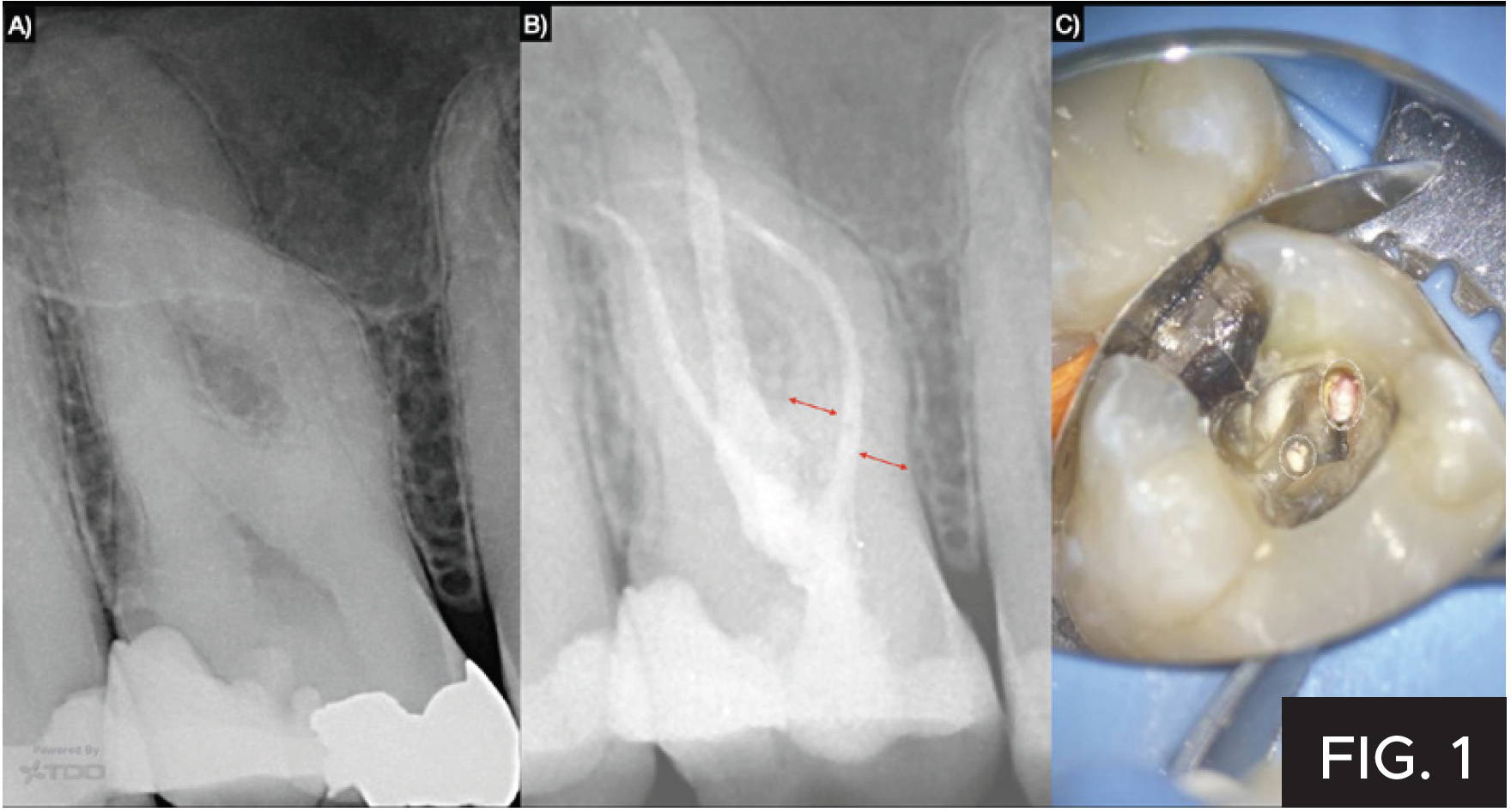
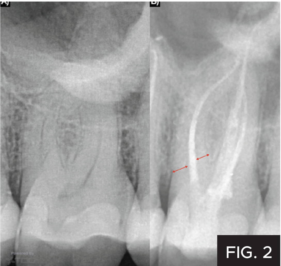
Directed dentin conservation refers to the preservation of valuable dentin structures and may play an important role in increasing the survival rate of endodontically treated teeth (1). Valuable dentin includes the pericervical dentin which refers to the root structure located 4 mm above and below the crest of bone (FIG. 1b) and (FIG. 2b) (2). Current best available evidence suggests that the main causes for extraction of root-filled teeth most often include recurrent caries or restorative and structural failures, with failures of true endodontic origin reported least frequently (3). Therefore, pericervical dentin conservation during access cavity preparation and instrumentation should be a priority of endodontic treatment.
Case Reports
Case #1 (FIG. 1)
Diagnosis: Pulp Necrosis with Symptomatic Apical Periodontitis
Working Length: 27 mm
Number of visits: two
Inter-appointment medication: Ultradent™ UltraCal™ XS 30%–35% Calcium Hydroxide Paste

OVER 175 YEARS OF DEDICATION
SS White®’s advancements are helping to create the masterpieces that are improving patient satisfaction and your bottom line. Few companies can trace a corporate history with a bedrock of innovation that spans back to 1844. Today, SS White Dental is a family owned and operated U.S. based business with more than 250 employees and worldwide distribution. Its corporate roots are found in the history of Samuel Stockton White, who began his career as an apprenticing dentist and ventured into his own business in 1844, manufacturing porcelain teeth in Philadelphia, Pennsylvania. The company proudly bears the name of its founder and continually maintains its original objective: better products for better dentistry.
SS White Endodontics files are the next generation of the minimally invasive RCT delivering a holistic strategy that focuses on conserving as much of the tooth as possible through minimally invasive dentistry.
Case #2 (FIG. 2)
Diagnosis: Pulp Necrosis with Symptomatic Apical Periodontitis
Working Length: 25 mm
Number of visits: two
Inter-appointment medication: Ultradent™ UltraCal™ XS 30%–35% Calcium Hydroxide Paste
Instruments used for access cavity preparation:
- 850-014M and 859-010M tapered diamond bur (SS White Dental)
- ESE014 safe end tapered carbide bur (SS White Dental)
- MB2 troughing: EndoGuide® Bur EG2 (SS White Dental)
Instrumentation Protocol
The coronal half of the canals were secured using a #10 K-file and sequentially enlarged in a crown down fashion using the SS White® DCTaperH™ #14/03, #17/04 and #20/06 rotary files with frequent recapitulation. Working length was determined with the electronic apex locator. After hand instrumentation to the full working length using a #10 K-file, the canals were shaped to their full length sequentially in a crown down fashion using the DCTaperH™ #14/03, #17/04 and #20/06 rotary files (SS White Dental), making sure to maintain patency and frequent irrigation in between file changes.
INSTRUMENTATION PROTOCOL
Full strength Sodium Hypochlorite with sonic activation.
OBTURATION TECHNIQUE
Custom fitted Fine-Fine (buccal canals) and medium-fine (palatal canal) gutta percha points with an epoxy based sealer and the warm condensation technique.
FINAL RESTORATION
Immediate restoration at the time of obturation was completed using Ultradent™ Ultra-Etch™ Etchant, Clearfil SE Protect bonding system (Kuraray) with dual cure activator and Luxacore (DMG-America).
Conclusion
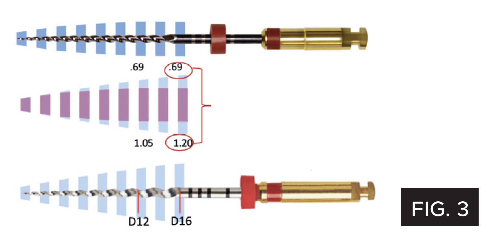
Safe instrumentation with pericervical dentin preservation is especially important in long and curved canals and is made possible with the regressive taper design and extremely flexible metallurgy of the DC Taper rotary file system (SS White Dental). The most popular rotary file systems have a diameter at the level of the canal orifice that approximates 1.2 mm. The corresponding DC Taper rotary file system has a diameter of 0.69 mm at the level of the orifice (FIG. 1c), allowing for maximal dentin conservation where it matters (FIG. 3).
REFERENCES
1. Clark D, Khademi J. Modern molar endodontic access and directed dentin conservation. Dent Clin North Am. 2010;54(2):249–73. doi:10.1016/j.cden.2010.01.001
2. Clark D, Khademi J, Herbranson E. The new science of strong endo teeth. Dentistry Today. 2013;32(4):112, 114, 116–7.
3. Nadeau B, Jung D, Vora V, Trends towards conservative endodontic treatment. Oral Health. 2019; May
About the Author

Bobby Nadeau, DDS, MSc
Dr. Nadeau is a registered Endodontic specialist in Canada, a Fellow of the Royal College of Dentists of Canada (FRCDC), and a member of the Canadian Academy of Endodontics. Dr. Nadeau’s specific interests in clinical endodontics include dynamic navigation for enhanced tooth tissue conservation during root canal treatment, probabilistic decision making, and focusing on patient-centered outcomes and restorative endodontics. He has authored and co-authored papers on various different aspects of Endodontics.
Discover More
This article was originally published in the Clinical Life™ magazine: Winter 2022 edition
Clinical Life™ magazine is a premier periodical publication by Clinical Research Dental Supplies & Services Inc. Discover compelling clinical cases from Canadian and US dental professionals, cutting-edge techniques, product insights, and continuing education events.


